Undergraduate students explore methods behind PROM1 retinal protein mutations through hands-on summer research program
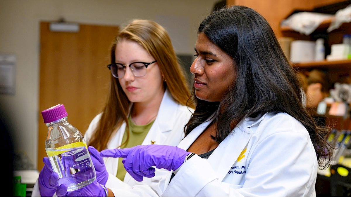
A summer research program hosted on the West Virginia University Health Sciences campus helped a pair of undergraduate students affirm their passion for vision research, as they explored how mutations of a particular retinal protein can lead to blindness.
Abigail Sheets, a junior biochemistry major at West Virginia University, and Karishma Beraj, a senior neuroscience major at the University of Pittsburgh, were two of the nine students who were accepted into the Summer Undergraduate Vision Research Fellowship Program in 2026.
The 10-week, full-time program provides undergraduate students from across the country with basic biochemistry and visual neuroscience research experience in areas exploring blinding diseases and new diagnostic and treatment methods. Each student enrolled in the program is paired with a faculty mentor on a small-scale, hands-on research project that they lead throughout the duration of the program.
Sheets and Beraj were each partnered with Professor and Vice Chair of Research at the WVU Eye Institute, Visvanathan Ramamurthy, PhD, on two separate projects that examined vision loss related to the mutation of a protein known as PROM1.
“Working with students through this program allows us to provide them with the type of extensive, hands-on laboratory experiences that are often unavailable to them during the regular academic year due to time and scheduling constraints, while they learn valuable skills and make meaningful contributions to major research studies here at West Virginia University,” Dr. Ramamurthy said.
Ramamurthy explained that PROM1 is a retinal protein that localizes on the outer segment of the light-detecting cells known as photoreceptors. When a mutation of this protein occurs, it can disrupt the formation of nascent discs in the retina and cause devastating vision loss.
Sheets’ project focused on replacing the damaged PROM1 gene with a paralogous gene known as PROM2 by injecting it into a laboratory model of the retina using a method known as adeno-associated virus (AAV) gene therapy and determining if it is successful in restoring vision and retinal function.
“PROM1 is found across the human body and is accompanied by PROM2 everywhere except for the retina,” Sheets explained. “This project allows us to explore why this protein is not naturally found in the retina and whether it can successfully replace a mutated PROM1 protein and protect against vision loss.”
Following successful AAV gene therapy, Sheets performed electroretinograms (ERGs) on the laboratory models, which were then sectioned, stained, and imaged, allowing her and the team to directly observe the structure of the retina and the number of photoreceptor cells that remained.
“It takes approximately one month for a virus introduced via AAV gene therapy to take effect, so during this time I was able to build my proficiency in all of the procedures required for data collection, such as performing dissections, slicing sections of eye cups, and plating and staining sections. Dr. Ramamurthy helped me with these tasks and allowed me to hone my skills by practicing on other laboratory samples. Once the time came to examine my own model, I was able to confidently complete every task, from performing the ERGs to image editing, all on my own,” she said.
While Sheets’ project explored treatment methods following the PROM1 gene mutation, Beraj’s project aimed to disrupt the mutation of the PROM1 protein entirely.
Beraj explained that her project hypothesized that inhibiting the mechanism by which the mutated PROM1 protein is degraded in the retina will allow the protein to stabilize and prevent further vision loss. Some of the laboratory techniques utilized for this research included western blotting to visually mark the PROM1 proteins in the retina, immunohistochemistry and immunofluorescence to determine the localization and prominence of the protein, and performing ERGs to compare photoreceptor responses to varying intensities of light stimuli.
Beraj said the sheer scope of this project has been an exciting challenge that has added some much-needed tools to her toolbelt as she looks ahead to the next steps in her educational journey.
“I’ve learned so much over the past several months thanks to this program and have been able to engross myself in this field even more than I imagined I would be able to,” Beraj said. “I’ve learned how to perform experiments, work with different pieces of laboratory equipment, perform imaging, but most importantly – I’ve learned what it means to find truly meaningful results and how to apply them. I look forward to applying this knowledge as I continue my education and seek out more lab experiences in the future.”
At the conclusion of the program in August, both Sheets and Beraj formally presented their research projects to their fellow classmates and faculty mentors during a student research symposium. Both students say they plan to become involved in more hands-on research opportunities as they continue their studies and work towards careers in medicine. Following the completion of their undergraduate studies, Sheets plans to attend optometry school, while Beraj aims to pursue medical school.
The Summer Undergraduate Vision Research Fellowship Program is hosted by the WVU Departments of Ophthalmology and Visual Sciences and Biochemistry and Molecular Medicine, and is led by Assistant Professors Michael Robichaux, PhD, and Saravanan Kolandaivelu, PhD. It is supported by the WVU Center of Biomedical Research Excellence (CoBRE), an initiative aimed at developing innovative methods to prevent, treat and slow the progression of blinding diseases. CoBRE is funded through a major grant from the National Institutes of Health.
To learn more about vision research at the WVU School of Medicine, visit medicine.hsc.wvu.edu/eye/research.