Submitting a Specimen
Kits are supplied by WVU. Kits include three vials:
- 10% formalin
- 3% glutaraldehyde in cacodylate buffer
- Michel's/Zeus immunofluorescent transport media
Once biopsies are obtained, place the tissue on a saline soaked gauze for evaluation with a dissecting microscope.
Evaluate for adequacy – presence of glomeruli, which appear as red dots if glomerular architecture is well preserved.
Submit the specimens (16 gauge needle cores) as follows:
- If three cores of renal parenchyma are obtained, put the first core directly into formalin and divide the other two cores according to the diagram below.

LM

EMIFEM
EMLMEM - If two cores of renal parenchyma are obtained, divide the cores up as shown in the diagram.
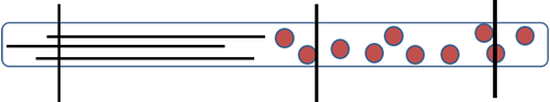
EMIFLMEM
EMLMIFEM - If only one core of renal parenchyma is obtained, the core must be divided up and placed into the three solutions. See the diagram below.

EMIFLMIFEM
Note: A minimum of two cores is recommended to have enough glomeruli for examination.
Address for overnight delivery
West Virginia University Hospital
Pathology Department – Renal/EM
Attention: Becky Radabaugh
1 Medical Center, Drive, Box 9203
Morgantown, WV 26506
Phone (304) 293-2092
Fax (304) 293-6249
Specimens are received Monday through Friday 8 a.m – 5 p.m. (excluding holidays).
UML Clients Contact
University Medical Laboratories
304-293-1030
888-865-3678