Follow the Tissue: Collection to Diagnosis
The following ran in the Daily Athenaeum as part of an inside look into what the Medical Labratory Science major training entails:
A case of Appendicitis
Appendicitis is inflammation of the appendix that usually results in emergency surgery to remove it. Have you ever wondered what happens to that tissue once it is removed?
Tissue removed surgically is sent to Anatomic Pathology where it is diagnosed by a pathologist. What most people don’t know is that Histotechnologists are the laboratory professionals who are responsible for preparing the tissue for the pathologist. This includes a series of complex processes.
The following five steps represent the standard process that every routine tissue specimen goes through for diagnosis.
Histotechnologists are essential for the appropriate handling and processing of tissue specimens. They are responsible for ensuring the quality of the processes, staining and testing so that the pathologist can make an accurate diagnosis.
Step one: Dissection
The appendix is dissected into small pieces for placement into a specimen case called a cassette. More complex specimens, such as cancerous tumors, are dissected by Pathologists’ Assistants. A description and dimensions of the appendix are documented for the pathologist. The cassette is placed in a preservative to prevent decay.
Step Two: Tissue Processing
The cassette is placed onto a tissue processor where the water in the appendix is removed and replaced with paraffin wax. The reason for this is to provide structural stability to the tissue. This process can take anywhere from 15 minutes to 10 hours depending on the type and size of the tissue.
Step Three: Embedding
The cassette is removed from the tissue processor and goes through embedding. A Histotechnologist transfers the appendix pieces from the cassette into a melted paraffin filled mold. The reason for this is enable sectioning. The paraffin is solidified and a tissue block is created.
Step Four: Sectioning
The tissue block is placed on an instrument called a microtome. The Histotechnologist uses the microtome to obtain paper thin sections of the appendix pieces. The sections are floated onto a warm waterbath and picked up onto a slide.
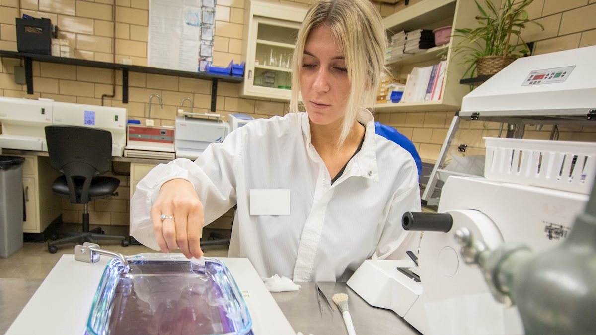
Step Five: Staining
The appendix pieces on the slide must be stained to identify tissue cells and structures. The routine stain performed on every tissue section is Hematoxylin and Eosin (H&E). A diagnosis of appendicitis can typically be made from an H&E; however, special stains may be performed if the pathologist suspects the presence of microorganisms or to verify the diagnosis.
Histotechnology is one of two tracks offered in the Bachelor of Science Medical Laboratory Science degree at West Virginia University.
The program provides an intensive combination of lectures, laboratories and clinical experience. For more information visit medicine.wvu.edu/medlabscience/.