Emerging Technologies
New Projects Currently Under Development by Our Group
Dedicated Head and Neck PET/CT Scanner
We are investigating the creation of a dedicated system to produce high resolution images of the head and neck region. This system will combine PET and CT imaging of the head and neck region to improve planning of radiation treatments for and the staging of head and neck cancers. Use of high resolution PET will facilitate accurate targeting of radiotherapy by supplying images of active tumor regions instead of bulk regions identified with anatomical methods. The PET images can also be used to more accurately assess local spread of the disease compared to current methods. The system will consist of two large, cooled, SiPM-based rotating scintillation detectors and a large FOV, cone beam CT scanner. The PET detectors are to be mounted on linear slides so that the distance separating them is adjustable. Therefore, the geometry of the PET scanner can be conformed to size of the patient and permits imaging down to the clavicle. The co-planar PET and CT scanner geometry will permit rapid co-registration of the images acquired from the two modalities. Simulation studies indicate that this scanner will have a spatial resolution of ~1mm FWHM. We are currently pursuing grant funds to continue development of this system.
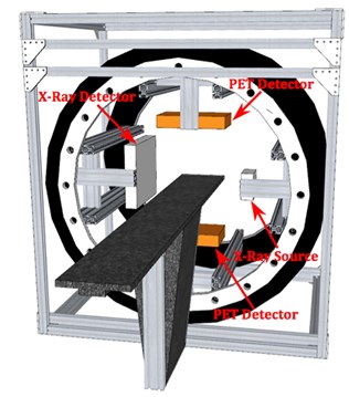
Schematic drawing of the dedicated head and neck PET/CT scanner.
Head and Neck Scanner Publications from our Group
- R.R. Raylman, A.V. Stolin, P. Sompali, N. Bunda-Randall, P.F. Martone, N.H. Clinthorne, Investigation of a Dedicated, High Resolution PET/CT Scanner for Staging and Treatment Planning of Head and Neck Cancer, IEEE Transactions on Nuclear Science, 2015;62(5):2057-2066.
TandemPET: A High Resolution PET Scanner
We are currently investigating development of a system for the PET imaging of small rodents, such as mice, for biomedical research purposes. This system will utilize a novel technology to produce a very high resolution PET scanner (known as TandemPET). High resolution is achieved by employing the dual detector variant of the virtual pinhole PET concept proposed by Tai, et al. The scanner consists of a small, high resolution detector (0.5x0.5x10mm^3 detector elements) with DOI measurement capability and a large detector with 1.5x1.5x10mm^3 detector elements. The two detectors rotate in a step-and-shoot fashion covering 360degrees. Simulation studies of the system using GATE revealed that, due to the very high intrinsic spatial resolution of the scanner (~0.5mm) it is necessary to apply correction for the effects of positron range (even for the relatively low energy positrons emitted by F-18). We have developed a three-dimensional, deconvolution method method for correction of this effect. To demonstrate the potential of the scanner for imaging the brains of mice, a simulated image of a mouse brain, using the MOBY digital phantom, was produced assuming use of FDG. After correction of positron range, it was possible to identify a number of very small brain structures (see image below). The imaging of mice is important to researchers developing new methods to detect and treat a number of disease such as numerous forms of cancer and neurological diseases (Alzheimer’s disease, for example). Due the use of a limited number of detector elements afforded by the dual detector geometry, the new scanner will be relatively inexpensive and compact, which is important for applications to the imaging of mice in research labs and pharmaceutical companies.
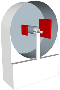
Schematic drawing of the TandemPET system showing the small, high-resolution detector and larger, lower resolution detector.
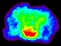
TandemPET image of the brain section of the MOBY phantom.
Tandem-PET Publications from our Group
- A.V. Stolin, S. Majewski, G. Jaliparthi, R.R. Raylman, Construction and Evaluation of a Prototype High Resolution, Silicon Photomultiplier-Based, Tandem Positron Emission Tomography System, IEEE Transactions on Nuclear Science, 2013;60(1):82-86. (NIHMSID 477184)