Intraoperative Beta Probe
The presence of residual disease following conservative surgical treatment is the most common cause of locally recurrent cancer. This phenomenon is primarily due to the desire on the part of the surgeon and patient to retain as much of the normal breast tissue and lymph nodes as possible. Thus great effort is taken to remove the minimum amount of tissue necessary to ensure a reasonable chance for effective treatment. Unfortunately it is very difficult for the surgeon to know how much tissue to excise, since it is usually hard to visually distinguish healthy from diseased breast tissue at the tumor margins. Furthermore, the standard conservative surgical protocol includes removal of most of the lymphatic system in the axilla ipsolateral to the breast containing the primary tumor, even though spread of the disease to this area is not known until after the excised tissue is examined. Removal of a large number of healthy lymph nodes can increase the morbidity and mortality associated with conservative breast cancer surgery. Although adjuvant radiation and chemotherapy is administered to reduce the likelihood of recurrent disease, if the amount of tumor burden left intact is too large, even this treatment will fail and subsequent surgery to remove additional breast tissue is necessary. In order to help reduce the amount of tumor left intact following the initial procedure and minimize the number of lymph nodes removed, we propose to design, construct and test a novel beta-sensitive surgical probe to intraoperatively detect areas of radiotracer-avid disease in situ. Due to the relatively short range of beta particles in tissue, techniques which rely on the detection of these emissions are potentially much more sensitive for localizing focal areas of cancer than are the more standard methods which require detection of highly penetrating gamma rays. The most unique aspects of this intraoperative probe system if the use of a dual, stacked solid-state detector unit.
The dual probe system utilizes ion-implanted silicon wafers to detect positrons emanating from by tracer-avid breast cancer cells and the background radiation caused by annihilation of the positrons. The two detectors are stacked. The front device detects both positrons (or electrons) and annihilation photons (or characteristic gamma emissions), the second device, located behind the first, detects only photons owing to the shielding effects of the first detector. Hence, signal from the second detector can be used to correct for photon contamination of the data obtained from the first device. Ion-implanted silicon detectors are used because of their very good energy resolution and noise characteristics compared to other solid-state detection devices, such as surface barrier detectors and plastic scintillators.
Instrumentation
The current version of the intraoperative beta probe is based on a compact, stacked, dual ion-implanted silicon detector unit designed at West Virginia University (United States Patent #5,744,805, #5,932,879 and #6,076,009)and Built by AMETEK, Inc.
The first detector detects both particulate emission and emitted from the source. Beta particles (positrons and electrons) emitted by most of the radionuclides used to label cancer-specific do not possess enough energy to be transmitted through the first wafer, therefore all the beta particle energy is deposited in the first detector. The second detector will thus only detect photons, and can hence be used as a monitor to estimate the photon contamination of the pure beta signal measured by the first detector. By subtracting the photon monitor's signal from the first detector's, the true beta count rate can be estimated.
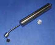
The detector unit as well as the preamplifier and amplifier units are housed in a stainless steel handle.
The amplified voltage pulses are relayed through a multi-wire cable to a signal processing unit where they are pulse-height analyzed; those signals above the threshold energy (typically 25 keV) are converted to TTL pulses. The TTL pulses are sent to a data acquisition card in the PCMCIA slot of a laptop computer. Both channels of data are counted and processed by the data acquisition software. Beta count rate is displayed on the computer interface. Additionally, the software can allow to the user to perform a statistical test of the data to estimate the significance of a positive reading. The data processing unit also contains the rechargeable battery pack used to power the system.
The amplified voltage pulses are relayed through a multi-wire cable to a signal processing unit where they are pulse-height analyzed; those signals above the threshold energy (typically 25 keV) are converted to TTL pulses. The TTL pulses are sent to a data acquisition card in the PCMCIA slot of a laptop computer. Both channels of data are counted and processed by the data acquisition software. Beta count rate is displayed on the computer interface. Additionally, the software can allow to the user to perform a statistical test of the data to estimate the significance of a positive reading. The data processing unit also contains the rechargeable battery pack used to power the system.
The project is current on hiatus. We are seeking translation opportunities to continue testing and commercialization efforts.
Acknowledgements
This work was supported by a research grant from the Whitaker Foundation.
Intraoperative Beta Probe papers published by our group
R.R. Raylman, R.L. Wahl, A Fiber-optically Coupled Positron-Sensitive Surgical Probe, Journal of Nuclear Medicine, 1994;35:909-913.
R. R. Raylman, S. J. Fisher, R. S. Brown, S. P. Ethier, R. L. Wahl, Fluorine-18- fluorodeoxyglucose-guided breast cancer surgery with a positron-sensitive probe: Validation in preclinical studies, Journal of Nucear Medicine, 1994;36:1869-1874.
R.R. Raylman, R. L. Wahl. Evaluation of Ion-Implanted-Silicon Detectors for Use in Intraoperative Positron Sensitive Probes. Medical Physics 1996;23:1889-1894.
R.R. Raylman, R.L. Wahl. Beta-Sensitive Intraoperative Probes Utilizing Dual, Stacked Ion-Implanted-Silicon Detectors: Proof of Principle, IEEE Transactions on Nuclear Science 1998;45:1730-1736.
R.R. Raylman. A Solid-State Intraoperative Beta Probe System, IEEE Transactions on Nuclear Science, 2000;47:1696-1703.
R.R. Raylman. Performance of a Dual, Solid-State Intraoperative Probe System with Fluorine-18, Technetium-99m and Indium-111. Journal of Nuclear Medicine 2001;42:352-360.
R.J. Lederman, R.R. Raylman, S.J. Fisher, P.V. Kison, H. San, E.G. Nabel, R.L. Wahl. Detection of Atherosclerosis Using a Novel Positron-Sensitive Probe and 18- Fluorodeoxyglucose (FDG), Nuclear Medicine Communications, 2001;22:747-753.