Visual Function and Morphology Core
The Visual Function & Morphology Core (VFM Core) provides equipment oversight, training, and assistance as needed to promote an environment conducive to success for the project leaders and others of the Visual Sciences Center of Excellence. Serving as a focal point for experimental design, data acquisition and functional analysis. In addition to equipment and expertise, the VFM Core offers technical assistance in transmission electron microscopy preparations, histology sample preparation, ERG/OKR assessments, and electroporation/orbital injections.
Equipment Available
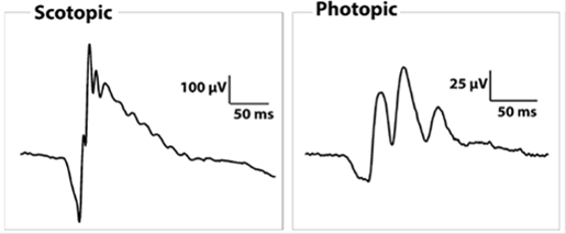
- LKC and Celeris Electroretinography (ERG) Systems
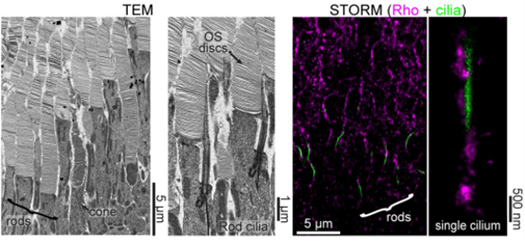
- JOEL Transmission Electron Microscope
- OptoMotry Optokinetic Response (OKR) System
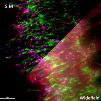
- SIM and STORM Super Resolution Microscopes
- Olympus Slide Scanner
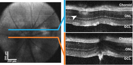
- Optical Coherence Tomography (OCT) System
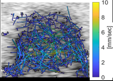
- Vevo Ultrasound System
- Zeiss and Nikon Confocal Microscopes
- Cryostat
- Vibratome & Ultramicrotome
- Ultramicrotome
- Image analysis workstation
ERG measurements of retinal function
Transmission electron and super resolution light microscopy of photoreceptor cell layer
Retinal blood flow tracing using microbubble contrast agent and the Vevo Ultrasound
OCT measurements of retinal thickness
Comparison of SIM super resolution to standard confocal microscopy